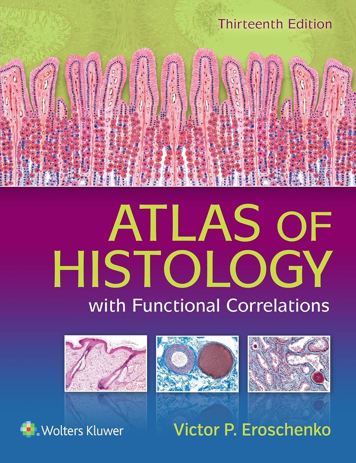
Struggling to connect histology structures with their real-world functions? You're not alone. Many medical and health science students find it challenging to translate textbook diagrams into actual tissue identification, especially when preparing for exams or clinical practice.
This 13th edition atlas solves that exact problem by pairing idealized histology illustrations with actual photomicrographs. You get the best of both worlds—clear, teaching-friendly diagrams alongside authentic tissue views that mirror what you'll see in the lab. This dual approach helps you develop the critical skill of recognizing structures in their natural state, not just in perfect textbook form.
The improved layout makes it easy to connect morphology with function, while the integrated Functional Correlation boxes throughout each chapter show you exactly how each structure works in the body. New photomicrographs and electron micrographs provide current clinical perspectives, and bulleted chapter summaries help you review efficiently before tests.
What sets this atlas apart is its emphasis on functional understanding. You'll learn not just what structures look like, but how they work and why they matter in health and disease. The additional histologic images and comprehensive review questions (375 in the book plus 250 online) give you multiple ways to reinforce your knowledge and track your progress.
Whether you're cramming for exams, preparing for lab work, or building your reference library, this atlas becomes your go-to resource. The combination of visual clarity, functional insights, and practical review tools makes histology less intimidating and more meaningful. It's the kind of reference that stays with you from your first anatomy course through clinical rotations and beyond.